Eva Reidemeister
Interviewer
Denis Poddubnyy
Rheumatologist
Long-form Interview:
This second part of the long-form interview turns to the management and treatment of Osteitis condensans. Denis Poddubnyy explains why the condition is challenging in clinical practice — and what conservative treatment options may help patients.
Eva Reidemeister: We’ve already touched on this a bit: But are there any guidelines for osteitis condensans yet? After all, the condition hasn’t been studied very well yet.
Denis Poddubnyy: Unfortunately, there is virtually nothing available on osteitis condensans ilii to date. There are some smaller studies, including one of our own, where we attempted a cross-sectional comparison of axial spondyloarthritis with osteitis condensans ilii. Despite the relatively small sample sizes, I believe we have identified the most relevant aspects, both clinically and in terms of imaging.
What we still don’t know is, first of all: What is the course of this disease? Are there self-limiting courses where the symptoms eventually subside, or do the symptoms persist? And: Is back pain always a part of it? And secondly, there are still no studies on how best to treat this disease and who is best suited to treat it.
In Germany, the situation is such that as soon as it is determined: This is not an inflammatory condition …
Eva Reidemeister: …you can really only administer painkillers.
Denis Poddubnyy: Yes, and rheumatologists are also losing interest in treating this condition. This is partly because rheumatologists are in short supply and are expected to focus on “true” inflammatory diseases. But seriously: These patients end up seeing orthopedists and are prescribed painkillers. But I think that, because bone marrow edema is often present, these cases require not only painkillers but also medications with both pain-relieving and anti-inflammatory effects, such as nonsteroidal anti-inflammatory drugs.
Eva Reidemeister: So, would those actually help?
Denis Poddubnyy: Yes, there are certainly situations where they do help. But on their own, they won’t improve the situation in the long term. The medications would, however, be important for managing symptoms so that the pain doesn’t become chronic. At the same time, though, you have to try to restore stability in the pelvis somehow, for example through physical therapy. Of course, performing physical therapy exercises regularly must not be accompanied by acute pain; that is, you must not make any movements that trigger acute pain. In that case, the combination most likely to be recommended to affected individuals is: physical therapy plus nonsteroidal anti-inflammatory drugs.
In addition, there are repeated attempts to improve stability in the sacroiliac joint through pelvic belts, through screw fixation, or through fusion of the sacroiliac joint. However, the studies on this are quite limited. And in my view, this would be the last resort for this condition—the very last thing one would consider. Experience shows that surgical intervention, in particular, does not solve all problems. I would always try to proceed with conservative measures first.
Eva Reidemeister: So we don’t have much information yet on the risk factors, but we also don’t know much about this disease in general. And one challenge is probably that the condition is simply very rare. As a result, there aren’t that many patients to study. Or where do the challenges in research lie?
Denis Poddubnyy: I think there are several factors at play here. Personally, I don’t believe that the disease is terribly rare . It has now become one of the most relevant differential diagnoses. But the challenge lies in distinguishing the diagnosis itself and the condition from axial spondyloarthritis. And the second challenge is: Who takes charge of this issue? Because here we find ourselves right at the intersection between orthopedics and rheumatology. And rheumatologists say: This isn’t inflammatory, so it doesn’t fall within our field. Orthopedists, of course, have other interests as well. So the affected patients are left somewhat adrift somewhere between two worlds.
But I think that because the condition is so relevant to rheumatologists [as a differential diagnosis, note], rheumatologists—and of course radiologists as well—need to initiate the appropriate studies. This includes long-term studies on disease progression and treatment efficacy. That would be very important. We need to convince potential sponsors that this is a significant condition that imposes a considerable burden on those affected. I am convinced of this. But we need more data to substantiate this claim,
Eva Reidemeister: Perhaps a somewhat technical question regarding research: At the beginning, you mentioned that the conditions are actually better identified on X-rays. That might be surprising, since X-ray technology is generally considered somewhat “old-fashioned” in imaging—MRI and CT are, after all, more modern. Does this amount to a case for why X-rays remain important?
Denis Poddubnyy: Yes, so if you know how to distinguish osteitis condensans from axial spondyloarthritis on an MRI, it’s not a problem. But I also believe that we have this problem of misdiagnosing osteitis condensans as axial SpA precisely because MRI is used more frequently and X-rays are no longer taken. With X-rays, it’s rather unlikely that bone marrow edema will be misdiagnosed as axial spondyloarthritis. With MRI, the likelihood is significantly higher that bone marrow edema will suddenly be misinterpreted as a manifestation of axial spondyloarthritis. However, X-ray technology has many drawbacks in the diagnosis of sacroiliitis and can also produce false-positive results.
Therefore, provided the resources are available, the trend is toward using cross-sectional imaging, such as MRI. We are also working to train radiologists and rheumatologists to distinguish between different processes in the sacroiliac joint to prevent misdiagnoses.
However, there are certainly countries where, for example, the healthcare system does not allow for an MRI scan to be performed right away. And that’s perfectly fine. It is entirely legitimate to start with an X-ray, and only if the findings are unclear or inconclusive is a referral made for cross-sectional imaging.
In fact, MRI is now the gold standard for the diagnosis or differential diagnosis of axial spondyloarthritis. It is important that the protocol includes a sequence sensitive to erosions so that these can truly be detected or ruled out. Even with the protocols used today, this is not always a given. We are also working to show rheumatologists and radiologists how important erosion-sensitive sequences are. This can sometimes also help avoid the need for a CT scan. In the past, CT was the gold standard for imaging structural changes in bone, particularly erosions. Meanwhile, MRI has caught up with CT and can essentially image bone changes just as well as CT.
I have also prepared a case study where various methods are applied. This is the case of a 45-year-old woman who comes to us from the orthopedics department with the question of whether she has ankylosing spondylitis. The suspicion stems from the fact that she suffers from long-standing back pain with inflammatory components. From the medical history, we know: There is a family history of ankylosing spondylitis, and she is HLA-B27 positive. She does not engage in physically demanding activities or hobbies, and she has two children.
At events where I’ve shown this X-ray, even experienced rheumatologists have said, “This is clearly a case of ankylosing spondylitis.” Because here you can’t see the joint space, so it’s ankylosis— and therefore axSpA.
That’s precisely the danger with X-rays: You can clearly see the triangular sclerosis here. But you can’t clearly see the joint space. That’s why you need cross-sectional images.
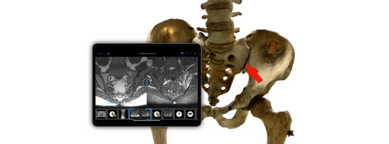
The case also includes a few cross-sectional MR images. STIR (this is the third series of our case) is a sequence optimized for fluid retention and inflammation, where you can see the bone marrow edema. In this sequence, however, you can see the structural changes. There is massive sclerosis. But it is impossible to say whether there is erosion or ankylosis present. The actual resolution is only achieved by using a sequence optimized for erosion, a so-called VIBE sequence, where one can see that the joint space is well preserved.
In this case, there is also further evidence that our theory regarding instability in the pelvic ring, specifically at the sacroiliac joint, is likely correct—and that is this capsular ossification. It occurs only when the joint is unstable and moves more than usual on a constant basis. Normally, there is only one to two millimeters of movement in this joint. If it increases, this is perceived as mechanical stress. This, in turn, leads to ossifications or bony spurs, similar to those seen in osteoarthritis. That is why it is now said that osteitis condensans is actually an extreme form of osteoarthritis. In the anterior, ventral region—where mechanical stress is greatest—both typical features of osteitis condensans ilii are visible: sclerosis and edema.
Eva Reidemeister: So we’ve come back to the terminology—with osteitis condensans being a type of osteoarthritis. In the case of ankylosing spondylitis, the nomenclature has completely changed in the meantime. We now refer to it as axial spondyloarthritis for various reasons. Could something similar happen here, where the disease might eventually be named something entirely different?
Denis Poddubnyy: Well, to be honest, I would have preferred to stick with the old terminology for axial spondyloarthritis [Editor’s note: i.e., Bechterew’s disease or ankylosing spondylitis]. There’s so much confusion. When you introduce new terminology, you really have to think very, very carefully about why. Well, in the case of axSpA, there were actually good reasons. The goal is to emphasize the inflammatory nature of the disease rather than its ankylosing nature. Here, I think if we stick with osteitis condensans, that would be fine. The main thing is that we understand what it is and can recognize it.
Eva Reidemeister: If that isn’t a fitting closing remark! Thank you very much, Denis Poddubnyy!
This interview with Dr. Denis Poddubnyy shows why Osteitis condensans ilii deserves much more attention as a relevant differential diagnosis of axial spondyloarthritis — not only in imaging, but also in clinical decision-making. Doctors who want to deepen their understanding can find further insights in the BCV Online Academy, Full Tier, with dedicated video lessons and curated case collections.