
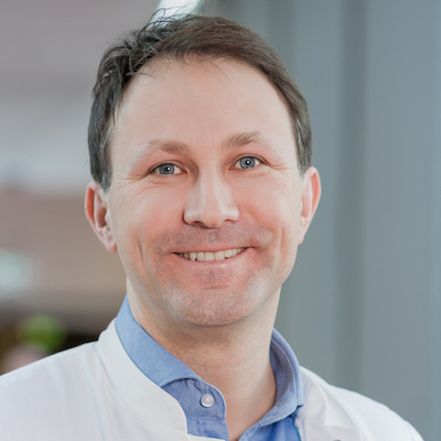
Dr. Hermann is a professor of radiology specialized in musculoskeletal imaging with a focus on arthritides. His research is dedicated mainly to the axial skeleton but the peripheral joints as well. The majority of his research projects are in the field of spondyloarthritis, where he is also connected with other researchers from Canada, UK, Germany, and Israel. He has also a strong track in applying innovative technology within medical education. He is an active member of ASAS, ESSR, DGMSR, and DRG and has won the Conrad Wilhelm Roentgen award in 2011, Felix Wachsmann award in 2014 and DVMB research prize in 2015. In 2019 he was promoted to associate professor.

You are currently viewing a placeholder content from Vimeo. To access the actual content, click the button below. Please note that doing so will share data with third-party providers.
More InformationYou are currently viewing a placeholder content from YouTube. To access the actual content, click the button below. Please note that doing so will share data with third-party providers.
More InformationYou need to load content from reCAPTCHA to submit the form. Please note that doing so will share data with third-party providers.
More InformationYou are currently viewing a placeholder content from Facebook. To access the actual content, click the button below. Please note that doing so will share data with third-party providers.
More InformationYou are currently viewing a placeholder content from MailerLite. To access the actual content, click the button below. Please note that doing so will share data with third-party providers.
More InformationYou are currently viewing a placeholder content from Instagram. To access the actual content, click the button below. Please note that doing so will share data with third-party providers.
More Information