Denis Poddubnyy
Rheumatologist
Osteitis condensans
When inflammation and bone sclerosis occur in the pelvic region near the sacroiliac joints, both rheumatoid conditions and bone changes—such as those caused by mechanical stress—are possible. In the differential diagnosis, axial spondyloarthritis (axSpA) and osteitis condensans ilii (OC) are therefore closely related.
In Germany, nearly one in three people regularly complain of back pain—in many cases, the symptoms are chronic.1 In about 10% of cases, the back pain is inflammatory.2 Imaging findings thus reveal inflammatory processes typical of inflammatory rheumatic diseases. A differential diagnosis that has received little attention to date but is important is osteitis condensans (OC)—better known in Germany as “hyperostosis triangularis ilii”. Inflammatory changes can also frequently be observed on imaging in OC. The condition, which can be viewed more as an extreme form of osteoarthritis, reminds us that inflammatory back pain is not automatically associated with inflammatory rheumatic diseases.
In a two-part interview, rheumatologist Denis Poddubnyy explains how osteitis condensans and axial spondyloarthritis (axSpA) can be reliably distinguished from one another.
New Terms for Spinal Disorders
“Ankylosing spondylitis” is considered an outdated term but remains in common use internationally. However, the term “ankylosing spondylitis” is more precise for severe cases involving irreversible functional limitations of the bones and joints, with “ankylosing” emphasizing the stiffness. Mild cases as well as the early stages of the same disease have also been referred to as non-radiographic axial spondyloarthritis (nr-axSpA) since the introduction of the ASAS classification criteria in 2009, because no structural changes are (yet) visible on conventional X-rays.
Is it axSpA or OC?
In axial spondyloarthritis, the term “spondylo” focuses on the vertebrae, while “arthritis,” as joint inflammation, indicates that we are dealing with a rheumatoid condition. Furthermore, the term “axial” tells us that we should consider the axial skeleton and surrounding structures, such as the sacroiliac joints.
Both the term “hyperostosis triangularis ilii,” commonly used in German-speaking countries, and the internationally more familiar term “osteitis condensans ilii” provide clues as to how we can recognize this condition. We are looking for inflammatory bone thickening (osteitis condensans) and should examine the ilium more closely. According to the German term, we find ossifications or bony outgrowths (hyperostosis) there, and these appear triangular in shape (triangularis) on the X-ray.
Denis Poddubnyy: However, we now know that this massive sclerosis, this bone densification, can occur in the sacrum just as it does in the ilium. It is simply easier to see it in the ilium than in the sacrum due to projection. And whether this sclerosis appears triangular on the X-ray image or not depends largely on which X-ray technique was used. If you take a conventional panoramic pelvic X-ray, it really does look like a triangle, just as described in the textbook.
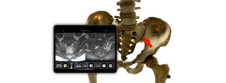
However, an X-ray is not always taken when axial spondyloarthritis or osteitis condensans ilii is suspected. Instead, magnetic resonance imaging (MRI) is frequently used.
Denis Poddubnyy: The difficulties begin with MRI, because osteitis condensans can be accompanied by quite pronounced bone marrow edema. This is what you don’t see on an X-ray, but what you can see very clearly on an MRI image. […]
For years, we have taught in rheumatology and radiology that bone marrow edema or osteitis—that is, active inflammation in the sacroiliac joint—is an early sign of ankylosing spondylitis. […]
The problem is not modern MRI technology itself, but rather how it is used.
Denis Poddubnyy: In fact, MRI is now the gold standard for the diagnosis or differential diagnosis of axial spondyloarthritis. It is important to use a sequence that is sensitive to erosions so that they can truly be detected or ruled out.
Clinical findings as clues
Clinical findings provide important clues for diagnosis. Negative results for the genetic marker HLA-B27 and normal levels of C-reactive protein (CRP) have differential diagnostic value and suggest osteitis condensans ilii rather than axial spondyloarthritis, but do not rule out axial spondyloarthritis on their own.
Stay in the loop by asking the right questions
Whether it’s an X-ray or an MRI—to interpret the findings correctly, radiologists need specific information from the patient’s medical history in addition to the imaging data. In addition to standard details such as age and gender, doctors should routinely ask about a family history of previous illnesses, according to the current S3 guideline.
Are there inflammatory rheumatic diseases such as axial spondyloarthritis in the immediate family? What other potential stress factors affect the axial skeleton? Does the affected person engage in physically demanding work or have a sporting hobby such as horseback riding? Being overweight also places additional strain on the joints and vertebrae. Another question to ask: Has the person ever been pregnant?
Osteitis condensans ilii:
Osteitis condensans ilii, a condition characterized by bone condensation and frequently inflammatory processes such as bone marrow edema, marks the intersection between orthopedics and rheumatology, with radiology playing a key role in diagnosis. Although not much is yet known about the pathogenesis of the disease, it is gaining importance in differential diagnosis. Denis Poddubnyy explains:
Denis Poddubnyy: Personally, I don’t believe the disease is terribly rare. It has now become one of the most relevant differential diagnoses. But the challenge lies in distinguishing the diagnosis itself and the disease from axial SpA.
Physicians from various specialties are called upon to collaborate even more closely in the future. Smaller studies have already identified important characteristics of the disease, but there are still unanswered questions:
- Are there self-limiting courses?
- Is deep-seated back pain always part of the clinical picture?
- What is the best way to treat osteitis condensans ilii?
Patients are often prescribed painkillers after diagnosis, yet the symptoms could potentially also be treated with nonsteroidal anti-inflammatory drugs (NSAIDs), as these have both analgesic and anti-inflammatory effects. This could prevent the pain experienced by OC patients from becoming chronic. Surgical intervention should only be considered once all other therapeutic options have been exhausted.
Furthermore, clinical studies could provide insight into additional treatment options. To what extent can physical therapy support the restoration of pelvic stability? Which physical therapy exercises are suitable for treating osteitis condensans ilii?
Summary
Osteitis condensans ilii is an important differential diagnosis for axial spondyloarthritis. The condition often occurs after one or more pregnancies as sclerosis of the parts of the ilium near the sacroiliac joint. On X-ray, OC appears as a triangular bone lesion. The typical profile of patients with osteitis condensans is as follows: a woman in her early 40s, likely with a history of one or two pregnancies. HLA-B27 test results are usually negative, and inflammatory markers are normal.
Our two-part interview series (part 1, part 2) with Dr. Denis Poddubnyy, conducted by Eva Reidemeister, shows why Osteitis condensans ilii deserves much more attention as a relevant differential diagnosis of axial spondyloarthritis — not only in imaging, but also in clinical decision-making. Doctors who want to deepen their understanding can find further insights in the BCV Online Academy, Full Tier, with dedicated video lessons and curated case collections.