Eva Reidemeister

Interviewer

Denis Poddubnyy

Rheumatologist

Long-form Interview:

Osteitis Condensans and the Pitfalls of Sacroiliac Joint Imaging (Part 1: Diagnosis)​

This long-form interview, conducted by Eva Reidemeister from Team BerlinCaseViewer, takes a deep dive into Osteitis condensans as an important but often misunderstood differential diagnosis of axial spondyloarthritis. Denis Poddubnyy explains why mechanical stress around the sacroiliac joint can mimic inflammatory disease on MRI — and how radiologists and rheumatologists can better distinguish the two.

Eva Reidemeister: I first tried to make sense of “osteitis condensans” by breaking down the term itself, probably because I’m a full-time writer. “Osteitis” basically means “inflammation of the bone,” and “condensans” means that something is densified. But what exactly is osteitis condensans, and how does the condition manifest itself?

Denis Poddubnyy: Well, osteitis condensans ilii—the correct term in its full form—is more of a historical term in this country, one that is actually used as a synonym for hyperostosis triangularis ilii. In contrast, osteitis condensans ilii is the term people around the world are familiar with.

In any case, it is a condition that, as is currently believed, has a mechanical origin. This means that mechanical forces act on specific areas of the sacroiliac joint, causing the bone in these areas to become denser to withstand increased mechanical stress.

Similar processes also occur in other joints. These are also referred to as osteoarthritis. For example, when cartilage in the knee joint is worn down  , the bone is suddenly exposed to increased mechanical stress and becomes denser. This so-called subchondral sclerosis is one of the main signs of osteoarthritis.

You can imagine a similar process occurring in the sacroiliac joints. However, in this case, the loss of cartilage is not the problem per se, but rather a certain mechanical instability in the joint, where excessive mobility can occur. The mechanical stress is greatest in certain areas, mainly in the anterior region—and that is where the bone also thickens.

This condition was already known in the past, particularly in women who have given birth. The reason is that during pregnancy, the pelvic ring loosens, creating new mechanical forces that were not present there before pregnancy.

As a result, certain areas near the sacroiliac joint become denser, and this can persist even after pregnancy and childbirth, especially if the integrity or necessary “stiffness” of the pelvic ring is not restored.

The condition was discovered and identified through an X-ray. And that is what makes X-ray technology so unique: this sclerosis—that is, osteitis condensans, a condition characterized by bone densification—was observed only in the ilium, specifically in the hip bone. This is also the origin of the term “osteitis condensans ilii.” And due to the projection, it looked like a triangle. That is where the term “hyperostosis triangularis” comes from.

However, we now know that this massive sclerosis, this bone densification, can occur in the sacrum just as it does in the ilium. It is simply that, due to projection, it is easier to see in the ilium than in the sacrum. And whether this sclerosis appears triangular on the X-ray or not depends largely on which X-ray technique was used. If you take a conventional pelvic overview X-ray, it really does look like a triangle, just as described in the textbook.

But if you use a special technique, such as a Ferguson view—which is a common technique for imaging sacroiliac joints when axial spondyloarthritis is suspected—then, when you use this technique, this sclerosis suddenly appears to migrate to the center of the joint and no longer looks triangular, but often like a circle or a round sclerosis.

Eva Reidemeister: So the technique used is crucial. That ties into my next question: Why is osteitis condensans ilii occasionally misdiagnosed as a rheumatic disease? Perhaps this also has to do with imaging, in that the condition then resembles other diseases, such as spondyloarthritides.

Denis Poddubnyy: Yes, that’s actually the point. So the reason this condition is occasionally confused with axial spondyloarthritis or ankylosing spondylitis is due to advancements in—or the imaging techniques commonly used today—specifically MRI. On an X-ray, it’s usually not confused with axSpA because it looks quite different.

There, you see massive sclerosis, and at the same time, the joint space is completely normal. That doesn’t happen with ankylosing spondylitis. So, in the case of ankylosing spondylitis, you always have sclerosis, erosions, and changes in the joint space, but here [in osteitis condensans ilii, note] it’s just massive sclerosis in a specific area of the joint.

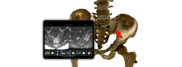
The challenges begin with MRI, because osteitis condensans can be accompanied by fairly pronounced bone marrow edema. This is something that isn’t visible on an X-ray but is clearly visible on an MRI scan. And for years, we have taught in rheumatology and radiology that bone marrow edema or osteitis—that is, active inflammation in the sacroiliac joint—is an early sign of ankylosing spondylitis. In fact, this edema can be quite pronounced in osteitis condensans and may also be accompanied by sclerosis, and then even occur alongside a further post-inflammatory change, namely the fatty degeneration of the bone marrow, known as fatty metaplasia.

These are likely natural processes that occur once bones have been damaged by bone marrow edema or inflammation. Then repair tissue, fatty infiltration, sets in, and this then progresses to sclerosis. And these three phenomena—bone marrow edema, fatty infiltration, and sclerosis—can be seen in the MRI image. Sometimes the findings are quite extensive, and even to the trained eye, this can resemble axial spondyloarthritis.

MR images of osteitis condensans (left) and sacroiliitis in axial spondyloarthritis (right) side by side. Both entities go along with bone marrow edema (green and blue arrows), but the location of their occurrence is slightly different.

Eva Reidemeister: But how can you tell them apart? It is, after all, an important differential diagnosis. We’ve discussed that I can recognize this triangular feature on an X-ray. But what if I start directly with an MRI image? Are there specific features there as well that would allow one to recognize osteitis condensans?

Denis Poddubnyy: You’ve prepared very well. You’re asking exactly the right questions, because we believe we can distinguish between them very well these days. I started by describing where the mechanical forces act most strongly on the sacroiliac joint—and that’s mainly in the anterior, ventral region. This means that if bone marrow edema is seen exclusively in this anterior, central region of the sacroiliac joint, it is reasonable to suspect that this bone marrow edema may be of mechanical—rather than inflammatory—origin. This is because inflammatory changes in axial spondyloarthritis are primarily seen in the middle region of the joint: where there is a good amount of cartilage—and not necessarily very far forward or very far back.

There are also structural changes that I did not mention when listing the changes associated with osteitis condensans, namely erosion—that is, the destructive alteration of bone. 

Inflammation in axial spondyloarthritis leads relatively quickly to the destruction of the endplate of the bone. This means that erosive changes, particularly in cases of severe inflammation, can be seen relatively early on. Sometimes you see a small erosion, a single lesion that may look like an erosion. This is, of course, nonspecific, but if you see multiple erosions in the sacroiliac joint, accompanied by bone marrow edema, it is more typical of an inflammatory disease and not of osteitis condensans ilii. 

In summary, we have two main characteristics: on the one hand, the location—at the very front in osteitis condensans and more centrally in the joint in axial SpA; and on the other hand, the presence or absence of erosive changes plays a very significant role. 

One more small point: The erosions in axial spondyloarthritis are not static. That is, they progress, and eventually, repair tissue forms after an erosion or within an erosion. This lesion is then called a “backfill,” meaning a filled erosion with scar or repair tissue. This is also something that is highly specific to axial spondyloarthritis and should not occur in the case of a mechanical problem, such as osteitis condensans—and, of course, no ankylosis either. Because that would essentially be the final stage of these processes: first destruction, then later repair leading to the complete closure of the joint space. This is only seen in axial spondyloarthritis. In osteitis condensans, in the vast majority of cases, the joint space is fairly well preserved. 

Eva Reidemeister: When I spoke with Joachim Sieper about axial spondyloarthritis, we discussed at length how radiology and rheumatology need to collaborate more effectively. Now the question here is: How do I recognize it in imaging? Are there differences? And to what extent do clinical values and the patient’s medical history play a role in the diagnosis? Perhaps that would steer the diagnosis in the right direction much more quickly.

Denis Poddubnyy: Yes, absolutely! That’s a very important question. Perhaps I should have started by saying that the specific clinical presentation is crucial. But there are always differences and variations, such as ankylosing spondylitis in women. And there are also cases of ankylosing spondylitis in women who already have children. 

The typical profile of patients with osteitis condensans looks like this: a woman in her early 40s, likely with a history of one or two pregnancies, often HLA-B27 negative, and with normal inflammatory markers. This would be a scenario where one should consider: Is it even axial spondyloarthritis? And if corresponding imaging findings also suggest that it is a mechanical problem, one would avoid diagnosing an inflammatory disease.

However, as I said, there are always exceptions: ankylosing spondylitis does occur in women. Ankylosing spondylitis does occur in HLA-B27-negative individuals, and ankylosing spondylitis does occur in people who do not have elevated inflammatory markers in their blood. That is why you have to consider both together: the clinical and the imaging information.

The duration is also important—that is, how long the symptoms have been present. This means that if someone comes to me and has had symptoms for 5, 6, or 7 years, and I see no erosive changes in the imaging or changes indicating past erosion, such as ankylosis or backfill, then I can say with a high degree of certainty that an inflammatory disease is rather unlikely.

With ankylosing spondylitis, one would expect certain changes typical of an inflammatory disease after 5, 6, or 7 years. Conversely, if the medical history indicates symptoms lasting several years and sclerosis is visible on the images—perhaps with bone marrow edema—but there are no destructive changes and no ankylosis, then a mechanical cause for these symptoms becomes likely.

Eva Reidemeister: We’ve already discussed prevalence and predisposition a bit. Osteitis condensans ilii is common in women who have already given birth. Are there other risk groups or population groups—perhaps age groups as well—where one should consider the possibility of osteitis condensans?

Denis Poddubnyy: Thank you for this important question as well. Because this also leads to the question: At what point do we say that osteitis condensans ilii is present? I believe it is still not well defined. In the past, when we only had X-ray technology, it could only be confirmed radiologically.

Today, we believe we can detect osteitis condensans even in its early stages. This gives us a broad spectrum of forms of osteitis condensans, since it all begins with bone marrow edema in the area exposed to mechanical stress. And I can very well imagine that only a portion of these edemas actually progresses to sclerosis at some point. This means that the range of findings that can be considered mechanically induced is significantly broader than in the classic X-ray image of osteitis condensans with pronounced sclerosis. 

And that is why our appeal to rheumatologists and radiologists is this: Always view bone marrow edema in the sacroiliac joint with great caution, and keep in mind the possible mechanical origin of these changes. In other words, the mechanical edema in osteitis condensans will primarily be localized anteriorly and will not be accompanied by destructive changes.

In addition to the wide range of changes that can occur, older people are also affected more frequently than younger ones. Furthermore, being overweight—that is, obesity—is a stress factor for the sacroiliac joints. This can lead to the development of bone marrow edema in the aforementioned area. It is important to note that bone marrow edema is a relatively nonspecific finding at the sacroiliac joint. This must always be considered within the clinical context and in the context of structural changes in the sacroiliac joint. 

We also assume that certain sports involving repeated stress or overloading of the sacroiliac joint, such as horseback riding or jogging, can also lead to bone marrow edema. 

Eva Reidemeister: Oh, although that’s a bit more dynamic! I would have thought of something like weightlifting.

Denis Poddubnyy: Actually, there was an interesting study on this by our Danish colleagues who examined marathon runners and ice hockey players using MRI a few years ago. 

They found bone marrow edema in about one-third of these otherwise healthy individuals. Bone marrow edema is a condition that can also occur physiologically, and the difference from the disease is this: in a healthy person, bone marrow edema develops and disappears again after a few days; in people who develop bone marrow edema associated with disease, the mechanical stress does not stop and is constant with every step. This then becomes symptomatic. Back pain develops, which, by the way, can also be inflammatory and does not stop spontaneously. 

Eva Reidemeister: That brings me to the next question: how patients—and conversely, doctors—can help prevent misdiagnoses through communication with one another. Because if, for example, I don’t mention a particular sport, it makes diagnosis more difficult. So what do both sides need to keep in mind: what questions should be asked, and what information should be shared?

Denis Poddubnyy: For the differential diagnosis, family history is also important—for example, whether someone in the family had an inflammatory rheumatic disease like ankylosing spondylitis. The types of sports, that is, the mechanical stress factors, are relevant. Most people don’t withhold this information, but doctors do need to actively ask about it. So we routinely inquire about mechanical stress factors, and the current version of the S3 guideline from the German Society of Radiology ( ) also recommends making this information available to radiologists. 

In other words, it is clearly stated there that information regarding pregnancies and specific stressors must be communicated to radiologists so that they can correctly interpret the imaging findings. When taking the medical history, we always inquire about the characteristics of the back pain. There is a symptom—or rather, a syndrome—of inflammatory back pain. This refers to back pain that improves with movement and worsens at rest, often occurs at night, particularly in the second half of the night, is accompanied by morning stiffness, and frequently changes in nature.

There is a common misconception here: People often equate the presence of inflammatory back pain with inflammation being the cause of that pain. But this is fundamentally incorrect. We really need to warn against this, because inflammatory back pain is merely a syndrome that can occur in various conditions, including osteitis condensans ilii, even though there is no actual inflammation present. There are also other conditions, such as degenerative spinal disease with active osteochondrosis and the development of a so-called Modic-1 lesion, where bone marrow edema forms in the spine around a damaged intervertebral disc. These patients come to us with typical inflammatory back pain, even though they do not have an inflammatory disease. 

So the presence of inflammatory back pain is always a very good starting point to investigate: Is there actually an inflammatory process present or not? But this says nothing about whether the inflammation is actually the cause of the back pain.  

Now that we have looked at how osteitis condensans can be recognized and distinguished on imaging, the second part of the interview moves on to the next key question: management and treatment.

Related posts:

[nextend_social_login login="1" link="0" unlink="0" heading=""]

Your feedback matters!
Let us know how we’re doing.